
SYSTEMATIC ANALISYS OF BONE TUMORS IN PLAIN RADIOGRAPHY

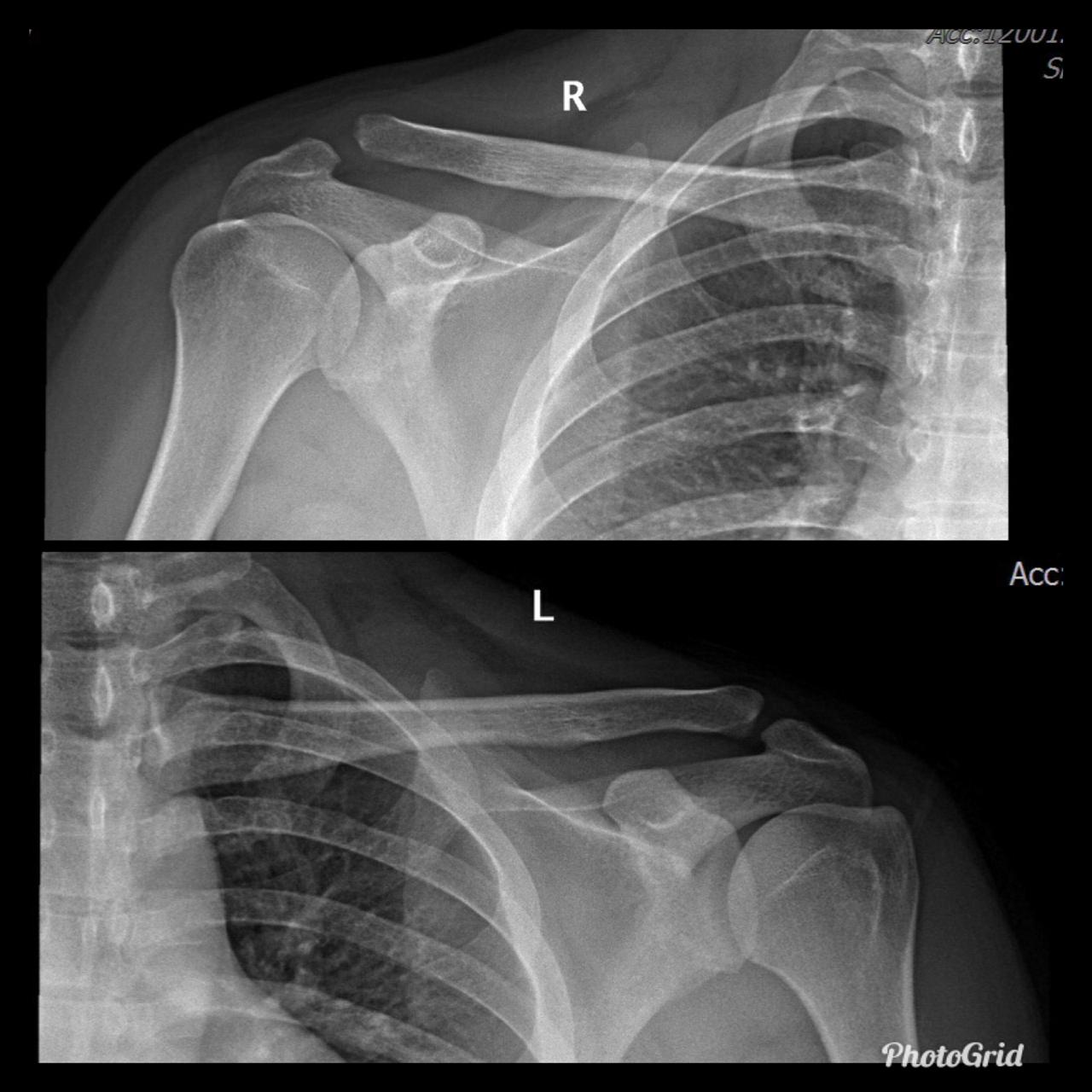


AC joint separation, often called a shoulder separation, is a dislocation of the clavicle from the acromion, with disruption of the acromioclavicular ligaments and/or coracoclavicular (CC) ligaments.
This injury is usually caused by a blow to the shoulder, or a fall in which the individual lands directly on the shoulder or an outstretched arm.
Treatment is immobilzation or surgical reconstruction depending on the degree of separation and ligament injury.
Rockwood Classification:
X-Ray Findings


Charcot joint, also known as a neuropathic joint or Charcot (neuro/osteo)arthropathy, refers to a progressive degenerative/destructive joint disorder in patients with abnormal pain sensation and proprioception.
In modern Western societies by far the most common cause of Charcot joints is diabetes mellitus, and therefore, the demographics of patients matches those of older diabetics.
Unlike septic arthritis, Charcot joints although swollen are normal temperature without elevated inflammatory markers. Importantly, they are painless.
Charcot joints are typically unilateral but are bilateral in ~20% (range 5.9-39.3%) of cases.
In the presented case there are destruction of talar head and neck with dislocation of talo-navicular joint, resorption of midfoot bones, subchondral sclerosis and multiple subchondral cysts on talocalcaneal aspect and cuboid facet of the calcaneum, progressive decrease of calcaneal inclination with typical rocker-bottom deformity, soft tissue swelling and arterial calcification. Bony debris are seen on dorsal aspect of the foot and posterior ankle joint. All findings are suggestive of Charcot neuro-osteoarthopathy, which is primarily an articular disease and most commonly located in the midfoot.

The fluid sign is one of the radiological features of osteoporotic fractures, and can be helpful in distinguishing them from metastatic vertebral fractures, as it is seen more often in osteoporotic fractures and is rarely seen in metastatic fractures.
The exact pathogenesis is not known, although proposed mechanisms include spontaneous avascular necrosis of the vertebral body (or Kümmell disease) or osteonecrosis at the site of an acute insufficiency vertebral fracture.
In fractured vertebral bodies, the fluid sign was adjacent to the fractured end plates and exhibited signal intensity isointense to that of cerebrospinal fluid on a background of diffuse hyperintensity in the vertebral body because of acute collapse.
It is seen in acute vertebral compression fractures that show bone marrow edema. In osteoporotic fractures, the fluid sign was significantly associated with fracture severity.


Titanium Elastic Nailing (TEN) is intended for fixation of diaphyseal fractures of long bones where the medullary canal is narrow or flexibility of the implant is paramount. The biomechanical principal of the Titanium elastic nailing is based on the symmetrical bracing action of two elastic nails inserted into the metaphysis, each of which bears against the inner bone at three points. The complication rates associated with Titanium elastic nailing have been reported to be minimal.


Osteomyelitis in diabetic foot is infection of the bone that 90% are results from contiguous spread of a skin ulcer. Consequently, the most common location for osteomyelitis is at the pressure points of the forefoot (metatarsal heads, IP joints) and in the hindfoot at the plantar aspect of the posterior calcaneus. Classic triad of osteomyelitis in plain radiograpy are lucencies, periosteal reaction, and bony destruction.
Unlike osteomyelitis, Charcot neuroarthropathy is primarily an articular disease and not related to overlying skin or soft tissue changes. Charcot neuroarthropathy commonly involves multiple midfoot bones. Features include joint instability, dislocation, destruction, disorganization, increased bone density, joint debris and deformity. Presence of subchondral cysts and intraarticular bodies with absence of the secondary signs for osteomyelitis support neuroarthropathy without infection.
In the early stage radiography will not demonstrate bone abnormalities, but MRI will show subchondral bone marrow edema. The subcutaneous soft tissues are not typically involved in Charcot arthropathy.
Osteomyelitis in chronic Charcot is usually located in the midfoot, while osteomyelitis in diabetic neuropathy without Charcot is usually in the forefoot and hindfoot.


Diabetic foot remains a challenge for the clinicians due to confusing clinical picture and associated complications. It may present as neuroarthropathy, septic arthritis, osteomyelitis, ischemic devitalised bone or as soft tissue complications such as cellulitis, myositis, ulceration, callus formation, sinus tracts, abscess, muscle denervation, tenosynovitis etc. Awareness of the various imaging findings of a diabetic foot, their relevance to the therapeutic decisions and correct usage of various imaging modalities to answer pertinent clinical questions are very important to improve the patient management and to reduce complications and morbidity.
Plain radiography is the preferred first line imaging investigation. It can show osseous structures and joint spaces well however it is neither sensitive nor specific. The detection rate and accuracy is low, especially in the scenario of early infection or neuroarthropathy, due to its lack of adequate demonstration of the soft tissues. In addition, Charcot’s foot and osteomyelitis may show overlapping radiographic features.
As foot infections in diabetic patients tend to be intractable, early introduction of therapy is paramount to prevent progression to gangrene and consequent amputation.


AP radiograph of the pelvis reveals extensive fluffy or "whiskering" enthesopathy of the iliac crests (red arrows), ischial tuberosities (yellow arrows), and the trochanters (black arrows). Note is also made of ossification of the iliolumbar ligament (blue arrow). Importantly, both sacroiliac joints appear normal. These findings are in keeping with diffuse idiopathic skeletal hyperostosis (DISH).
Diffuse Idiopathic skeletal hyperostosis (DISH) is a bone-forming diathesis primarily affecting the spine, with ossification of tendons and ligaments. Most of us are familiar with the spinal findings; however there are extraspinal manifestations as well such as hyperostosis at ligament attachments in the pelvis, calcaneus, tarsal bones, ulnar olecranon and patella.
Usually these are incidental findings without significant morbidity.


A Tillaux fracture is a traumatic Salter–Harris type III fracture through the anterolateral aspect of the distal tibial epiphysis, with variable amounts of displacement.
It accounts for 3-5% of pediatric ankle fractures and more commonly seen in girls. Tillaux feacture is seen in children nearing skeletal maturity (12-14 years old), when the medial epiphysis had closed but before the lateral side has done so.
The fracture commonly results from an abduction-external rotation force, causing the anterior tibiofibular ligament to avulse the anterolateral corner of the distal tibial epiphysis, at the opposite end to a Wagstaffe-Le Fort avulsion fracture, resulting in a Salter Harris Type III fracture.
Variability in fracture pattern is due to progression of physeal closure as anterolateral part of distal tibial physis is the last to close. When the lateral physis is the only portion not fused, external rotation may lead to Tillaux or Triplane fractures. Lack of coronal plane fracture in the posterior distal tibial metaphysis distinguishes this fracture from a triplane fracture.
Associated conditions commonly seen with this fracture are distal fibular fracture (usually SH I or II) and ipsilateral tibial shaft fracture.
If the displacement at fracture is less than 2 mm, it may be managed conservatively. However, displacement requires open reduction and internal fixation, especially when displacement is over 2 mm.
As with any intra-articular fracture if a step is left in the articular surface, then the joint will go on to premature secondary osteoarthritis.
This fracture pattern is named after Paul Jules Tillaux, a French Anatomist and Surgeon (1834-1904).

Pediatric traumatic hip dislocation are usually posterior and may occur due to low energy sports injuries in children less than 10 years of age. Hip dislocation are more common than hip fracture in pediatric patients and 80% are traumatic posterior dislocations.
Most of these cases can be diagnosed on AP pelvis films, which show loss of congruence of femoral head with acetabulum. Lateral view is sometimes is used to differentiate between anterior vs. posterior dislocation and to scrutinize femoral neck to rule out fracture prior to attempting closed reduction.
Treatment is urgent closed reduction under general anesthesia or sedation. Open reduction may be required if there is an intraarticular fragment following reduction. Post reduction imagings are necessary to inspect for joint incongruity or nonconcentric reduction.
MRI is a study of choice for any abnormal findings on post-reduction radiographs such as joint widening. In MR study, in inspect for joint incongruity or nonconcentric reduction. Entrapped labrum or capsule is best evaluated via MRI.
CT is second choice behind MRI for post-reduction evaluation, and also radiation exposure should be considered. Osteochondral fragments can be seen in older children and are easily detected by CT.
Delayed in reduction can lead to complications such as osteonecrosis, coxa magna, redislocation or nerve injury.


Distal clavicle osteolysis is a unique disease most likely due to an overuse phenomenon. Distal clavicular osteolysis (DCO) follows both chronic repetitive stress and single acromioclavicular trauma. Acute distal clavicular osteolysis was first described in 1936.
The exact aetiopathogenesis is unclear, but AC-joint trauma and subchondral microfractures seem to be involved. Subsequent attempts at repair are insufficient and the final result is osteolysis. It is unclear why changes predominate in the distal clavicle while the acromion is relatively spared.)
Mostly affected males in their 20s and commonly seen in weightlifters, symptoms usually begin with an insidious aching pain in the AC region that is exacerbated by weight training. Clinical findings are often nonspecific and frequently overlap with those of labral or rotator cuff tears. On examination, patients have point tenderness over the affected AC joint and pain with a cross-body adduction maneuver.
Conventional radiographs may remain normal during the first months or years. A 15° cephalad inclination avoids superposition of the scapular spine with the AC-joint (Zanca view).
Radiographic changes include cortical thinning, irregularity and microcysts in distal clavicle and mild AC–joint widening. A late finding is tapering of the distal clavicle.
MRI is far more sensitive to detect DCO in an early stage. The most common MR-finding is bone marrow oedema in the distal clavicle, sometimes also in the articular part of the acromion, but less distinct.
Often, a hypointense line is seen in this area of clavicular bone marrow oedema, representing a subchondral fracture. AC-joint abnormalities are common, and include effusion, mild widening, intra-articular bone fragments and capsule hypertrophy. Other MR-findings are similar to those seen on radiographs, as described above.
Bone scan may also shows increased uptake in the distal clavicle, which could be seen earlier than radiographic changes.
Treatment is essentially conservative, consisting of rest and nonsteroidal anti-inflammatory drugs (NSAID's), and is usually successful. In severe cases, resection of the distal clavicle is indicated. If left untreated, the process may cause progressive resorption of lateral aspect of the clavicle, erosions and cupping of the acromion and dystrophic calcifications.
DCO should be considered in the differential diagnosis of shoulder pain in the appropriate population. Therefore, analysis of MR arthrographic studies of the shoulder should not be restricted to evaluation of the rotator cuff and capsulolabral system, but the AC-joint should be scrutinised as well.


Trigger finger is a type of stenosing tenosynovitis. It develops due to repetitive microinjury from frequent flexion-extension movements of the fingers.
Primary trigger finger occurs most commonly in the middle fifth to sixth decades of life and up to 6 times more frequently in women than men.
A patient with trigger finger finds it difficult to straighten or bend the affected finger. The finger transiently gets locked in the flexed position and with a painful snapping sensation goes into extension.
Thickening and hyper-vascularization of the A1 pulley are the hallmarks of trigger fingers on sonography. Other frequently observed features include distal flexor tendinosis and tenosynovitis.
The first annular pulley (A1) at the metacarpal head is by far the most often affected pulley in trigger finger, though cases of triggering have been reported at the second and third annular pulleys (A2 and A3, respectively), as well as the palmar aponeurosis.
The level of thickening can be variable with some authors suggesting the normal value being around 0.5 mm with thickening suggested when the diameter is over 1.1 mm.
The condition can be conservatively managed with splinting, NSAIDs, and local steroid injections or may require a surgical section of the A1 pulley when the pulley is markedly thickened.


Olecranon bursitis refers to inflammation of the olecranon bursa. The olecranon bursa is a subcutaneous space lined with a synovial membrane that secretes fluid to provide smooth and almost frictionless motion between the skin, the subcutaneous tissues, and the olecranon. Because of its superficial location, it is a common site for injury, inflammation, and infection. Repeated traumatization of the elbows at work led to common terms for different forms of occupational bursitis such as “student's elbow” or “miner's elbow”.
Approximately one third of the cases of olecranon bursitis are septic. Many patients with septic olecranon bursitis lack a history of trauma or a visible injury over the olecranon.
Lateral radiograph of the elbow reveals soft tissue swelling superficial to the olecranon. Ultrasound may show a fluid collection in the olecranon bursa, features of synovial proliferation and/or hyperemia.
CT will show fluid density at the subcutaneous tissue superficial to the elbow. Bursal fluid collection in MRI has the following features: hypointense-T1, mainly hyperintense-T2, and enhancement of bursal margins in post contrast imaging.


Acute gouty arthritis of the first metatarsophalangeal joint, termed “podagra,” was first identified by Egyptians in 2640 B.C. and continues to be a medical health problem today. The hallmark of gout is hyperuricemia with subsequent deposition of monosodium urate (MSU) crystals, which leads to inflammation and symptoms. Gout commonly involves specific joints and anatomic structures, and knowledge of these sites and imaging appearances are clues to the correct diagnosis.
Although diagnosing gout generally is straightforward, atypical disease may present a challenge if it is associated with unusual symptoms or sites, discordant serum urate level, or mimics of gout. Dual-energy computed tomography (CT) may be used to differentiate urate crystals from calcium by using specific attenuation characteristics, which may help diagnose gout. In patients with known tophaceous gout, dual-energy CT may be used for serial volumetric quantification of subclinical tophi to evaluate response to treatment.
Dual-energy CT can quantitatively identify monosodium urate crystal deposits with high sensitivity and specificity within joints, tendons, and periarticular soft tissues.
Given the utility of dual-energy CT in challenging cases and its ability to provide an objective outcomes measure in patients with tophaceous gout, dual-energy CT promises to be a unique and clinically relevant modality in the diagnosis and management of gout.


Medial tibial stress syndrome (MTSS), also known as shin splints, describes a spectrum of stress injury that occurs at the medial tibia. This term is often used to indicate any type of tibial stress injury or the earlier manifestations of a tibial stress lesion before a fracture component can be identified. It is considered a low risk stress fracture.
Shin splints are a common exercise-related problem. Typically occurs in athletes (e.g. runners/jumpers) and is characterized by localized pain that occurs during exercise at the medial surface of the distal two-thirds of the tibial shaft, where muscles attach to the bone. Shin splints often occur after sudden changes in physical activity. These can be changes in frequency, duration and intensity.
Several conditions can cause shin pain, including stress fractures, tendinitis, and chronic exertional compartment syndrome. Other factors that contribute to shin splints include having flat feet or abnormally rigid arches and exercising with improper or worn-out footwear.
A "one-leg hop test" is a functional test, that can be used to distinguish between MTSS and a stress fracture : a patient with MTSS can hop at least 10 times on the affected leg where a patient with a stress fracture cannot hop without severe pain.
MRI is the most sensitive radiological examination (~88%). It may demonstrate a spectrum of findings ranging from normal to periosteal fluid to marrow edema to actual stress fracture. The medial cortex (+/- posterior cortex) is most commonly affected.
Simple measures can relieve the pain of shin splints. Rest, ice, and stretching often help. Taking care not to overdo your exercise routine will help prevent shin splints from coming back.


Avulsion fractures of the calcaneal tuberosity are uncommon, accounting for 1.3–2.7% of all calcaneal fractures.
The calcaneus is the primary weight bearing bone in the heel, and its many surface contours render it a relatively difficult bone to visualize in its entirety. The stabilizing ligaments that hold the calcaneus in place occupy very specific locations, and the Achilles tendon enthesis is in a relatively constant location; therefore, avulsion fractures occur in reproducible locations.
Fractures of the tuberosity are either from an avulsion or shear-compression mechanism of injury, with the latter constituting most fractures. Avulsion may occur from sudden tension on the Achilles tendon from falling on a plantarflexed foot when the calf muscles are actively contracted, hyperextension of the ankle, or while pushing off a dorsiflexed foot such as in a sprinter beginning a race.
There are four types of avulsion fractures: type 1, simple avulsion with a variable-sized bone fragment; type 2, beak fracture with a horizontal fracture extending into the posterior body; type 3, infrabursal avulsion by the superficial fibers of the middle third of the Achilles tendon; and type 4, small beak fracture avulsed from the deep fibers of the tendon.
An important pitfall is a neuropathic avulsion fracture of the tuberosity in a patient with long-term diabetes mellitus. In these patients, the fracture occurs without a history of significant trauma or overuse activity. The primary fracture line is parallel to the apophyseal scar, and the fracture affects the superior cortex but not always the inferior cortex.
The fracture also tends to extend posteriorly with a horizontal component immediately distal to the enthesis of the Achilles tendon. When the fracture is imaged sequentially, distraction and fragmentation are common later findings. Neuropathic fractures are important because they have a much higher incidence of infection, nonunion, malunion, and failure of fixation and require a much longer time to heal than nonneuropathic insufficiency fractures.
Another pitfall occurs in children. A Salter 3 fracture of the apophysis in a skeletally immature patient may mimic Sever disease when it is not significantly displaced. A systematic evaluation of the calcaneus with attention to areas of vulnerability will assist those who interpret ankle and foot radiographs in maintaining a high diagnostic accuracy for these fractures.
Typically, treatment is with open reduction-internal fixation (ORIF), although minimally displaced fractures may be treated with closed reduction.


Congenital pseudarthrosis of the tibia (CPT) refers to nonunion of a tibial fracture that develops spontaneously or after a minor trauma. It is a rare pathology, occurring in only 1 out of 250,000 births. The cause of CPT is currently unknown; however, there is a strong association with neurofibromatosis in 50% of cases and an association with fibrous dysplasia in 10% of cases. The pseudarthrosis usually develops within the first two years of life; but may be undetected up to the age of 12 years. Bilateral occurence is rare; the fibula is affected in one third of the patients.
Congenital tibial pseudoarthrosis is characterized by segmental osseous weakness, resulting in anterolateral angulation of the bone. Reports indicate the pathologic processes of CPT are linked to the periosteum, which forms the outer layer of bones and is crucial for regrowth following a fracture. Patients with CPT have a thick scar layer surrounding their bones rather than the periosteum. For this reason, once a fracture has occurred it will not heal spontaneously.
Classification systems describing prognostic radiographic characteristics, natural history, onset, mobility, therapeutic guidelines, and variables influencing the outcomes have been proposed by many different authors. Three classifications of CPT are commonly used. The Andersen classification differentiates the morphology of pseudoarthrosis as dysplastic, cystic, or sclerotic types, in addition to a clubfoot type that arises because of accompanying abnormalities. Crawford described four types of CPT, all types have in common an anterolateral bowing of the affected tibia. The classification of pseudoarthrosis by Boyd consists of six types and includes a type V, which is characterized by a complementary dysplastic fibula and a type VI, in which an intraosseous neurofibroma or schwannoma is evident.
While many classification systems exists, the most important criterion is the status of the tibia (fractured versus intact). Treatment is mainly surgical, with bracing used to supplement surgical procedures.

Knee bursae are sacs surrounding the knee joint that are filled with synovial fluid.
They facilitate movement and reduce friction where tendons or muscles pass over bony prominences.
The knee bursae can be either communicating or non-communicating with the knee joint itself.
There are four bursae anterior to the knee joint, each two bursae at medial and lateral knee, and also four bursae posterior to the knee joint.